Meningioma
Meningioma is a type of brain tumor that is usually considered benign. These tumors originate in the lining of the brain, rather than spreading (metastatic) from any other organ of the body. Meningioma is one of the most common brain tumors. Meningiomas are usually not adherent to brain tissue. This plays an important role in removing the entire tumor during surgery.
Meningiomas can be detected asymptomatically depending on their location. In such cases, the location and size of the tumor can be monitored according to the general clinical condition of the patient. In patients who present with symptoms, especially those with a pronounced edema effect and tumor size of more than 3 cm, it is aimed to treat the tumor by microsurgical removal.
Meningiomas can be defined in different ways according to their histopathologic type, location in the skull and relationship with the surrounding vasculature. They are most commonly seen in the upper part of the skull and the front of the head (anterior skull base).
- Convexity Meningiomas: Occurs at the top of the head.
- Parasagittal Meningiomas: Occurs in the upper middle part of the skull. The relationship with the sagittal sinus (one of the most important vascular systems of the brain) is very important in tumors of this region. According to the relationship of the tumors of this region with the sagittal sinus, the "Sindau Classification" defines the location and condition of the tumor more clearly.
- Posterior Fossa Meningiomas: Occurs at the back of the head.
- Tentorial Meningiomas: It originates in the deep inner part of the brain called tentorium.
- Cerebellopontine Corner Meningiomas: It is the type seen in the posterior outer part of the skull, in the area at the junction of the brain-cerebellum-brainstem regions. Especially the relationship with the auditory nerve and facial nerve plays a critical role.
- Intra-Orbital Meningiomas: It is caused by the eyeballs.
- Olfactory Groove Meningiomas: It occurs in the brain at the front of the skull in the area where the olfactory nerve passes.
- Spinal Meningiomas: It originates from the spinal nerve.
Symptoms of Meningioma in the Brain
Although meningiomas are defined as "benign tumors", their slow growth and late onset of symptoms cause the treatment option to be microsurgical at the time of diagnosis. For this reason at first they are asymptomatic. It may be detected incidentally by CT or MRI after trauma. The most common symptom is severe and persistent headache, but there may also be symptoms such as loss of speech, difficulty in movement, memory loss, depression, and urinary incontinence, which can seriously impair quality of life.
Symptoms of meningiomas are as follows:
- Headache, dizziness
- KIBAS (symptoms due to increased intracranial pressure)
- Weakness and instability in the arms and legs
- Personality change, behavioral disturbance, maladjustment, depression
- Loss of strength and sensation
- Epilepsy (Seizure)
- Visual impairment
- Nerve paralysis
- Impaired sense of smell
What causes meningioma?
Although the exact causes of brain tumors have not been fully determined, some risk factors are known to be effective. Brain tumors can occur at any age, but the incidence increases with increasing age. Genetic predisposition plays an important role in the development of brain tumors; individuals with a family history of brain tumors have an increased risk compared to healthy individuals. Some genetic diseases increase the likelihood of developing brain tumors. Brain tumors are more common in Caucasians than in blacks. Exposure to high doses of radiation and certain chemicals also increases the risk.
Diagnosis
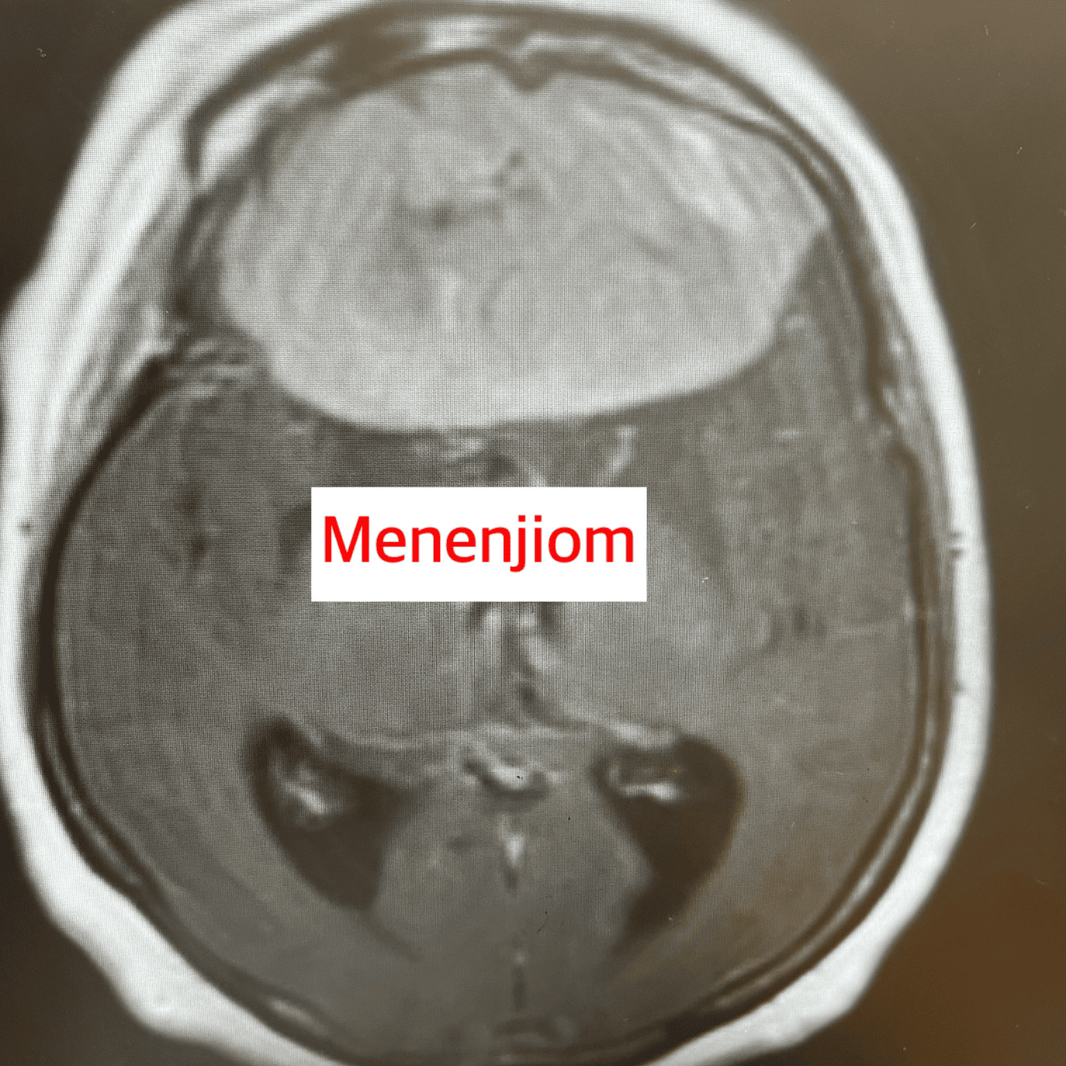
Meningiomas are detected by MRI and CT scans. In cases of suspected tumor, contrast-enhanced MRI is performed to examine radiological parameters such as the exact size of the tumor, its relationship with surrounding tissue, and the effect of tumor-related "edema". However, for a definitive diagnosis, that is, for the exact diagnosis and determination of the type of tumor, the tumor must be removed and sent to pathology histopathological diagnosismust be put in place.
Meningioma Surgery and Treatment
We have three main options for meningioma treatment: observation, surgery and radiosurgery. A number of components such as the patient's general health status, age, size and location of the tumor, and the extent to which it affects quality of life are taken into consideration and the patient's meningioma treatment is planned. The treatment journey is tailored to each patient's specific situation.
Observation
When meningiomas are detected, if the tumor is small, does not cause brain edema and does not show any clinical symptoms, monitoring without treatment may be an important option. These patients are called for follow-up at regular intervals and the tumor status is regularly checked by magnetic resonance imaging (MRI). However, this observation strategy is only applicable to some groups of patients. If the tumor is small, does not compromise brain function and does not affect the patient's quality of life, only monitoring can be planned instead of treatment. This approach may also be preferable in patients for whom surgery poses a high risk and in elderly individuals with no obvious symptoms.
Meningioma Surgery
Meningioma surgery is the most common method in the treatment of meningiomas. The tumor is removed completely or as much as possible with a microsurgical approach. As mentioned in the introduction, there is a distinct interface between benign tumors and the brain. This makes it possible to remove them completely by surgery. The vast majority of meningiomas are benign.
If the tumor reaches a certain size, touches critical nerve tissues, reduces the quality of life of the person, and is life-threatening, surgical treatment of meningiomas is necessary.
Under general anesthesia, after the appropriate surgical position is given according to the location of the tumor, the head is immobilized and fixed with a spiked headgear. Then the neuronavigation system, the 3-dimensional structure of the tumor is determined during surgery. After sterile staining and covering, the tumor is accessed by following the appropriate surgical rules. Some tumors may be associated with critical nerve structures depending on their location. In the planning of these cases neuromonitor may need to be used. The aim of meningioma surgery (although there are exceptions) is to remove the entire tumor.
Whether there is a recurrence after meningioma surgery, i.e. whether the tumor will recur or not, varies according to parameters such as the detailed pathological analysis of the meningioma and how much tumor was removed.